A-Z OF SKIN
Basal Cell Carcinoma (BCC)
BACK TO A-Z SEARCHLast updated: December 2023
Also known as… a Rodent Ulcer
What is basal cell carcinoma?
Basal Cell Carcinoma (BCC) is the most common type of skin cancer. BCC accounts for more than three-quarters (75%) of all skin cancers. 1
Most basal cell carcinomas are slow-growing and almost never spread to other parts of the body. However, if they are left untreated, they can damage or destroy the skin and surrounding tissues and cause an ulcer known as a rodent ulcer.
Who gets basal cell carcinoma?
Individuals who are at a greater risk of developing a basal cell carcinoma include those with fair skin, with a strong family history of BCC, and with a Celtic background.
What causes basal cell carcinoma?
There are many factors that lead to the development of most cancers however sun exposure is by far the most important factor in the development of BCCs. This excessive sun exposure usually occurred many years before the BCC develops.
What does basal cell carcinoma look like?
BCCs usually start with a subtle change on the skin, often a small bump or a flat red patch. BCCs develop very slowly over months and years, steadily becoming larger and more obvious. Eventually they may appear as a non-healing sore. BCCs are often not noticed until relatively well developed and their appearance can be confused with that of a naevus (mole), patch of dermatitis or a scar. A lesion that bleeds on the face is quite suspicious of BCC.
- Superficial
Superficial BCCs are confined to the very top layers of the skin so are often more easily treated with topical and non-surgical techniques. They can however become relatively broad in size. Superficial BCCs are especially common on the trunk.
- Nodular
This type of BCC usually appears as a rounded lump on the skin. They are less likely to have invisible extension under the surface of the skin than other types of BCC.
- Infiltrating
These are the most troublesome type of BCC as they are usually larger than they appear to the naked eye. They are usually the most difficult to see and are often not detected until well advanced. It is therefore not uncommon for this type to be inadvertently incompletely removed with treatment, allowing the BCC to continue to grow and recur. Morphoeic and micronodular are different types of infiltrating BCCs.
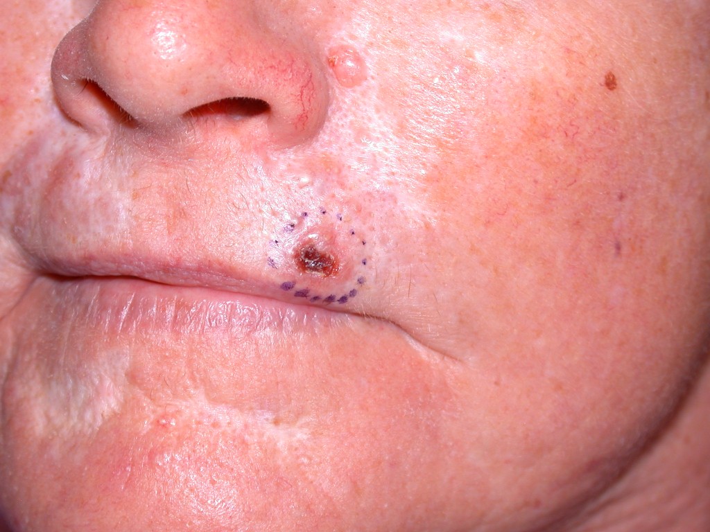
Figure 1. BCC on the lip. Image reproduced with permission of Dr Chris Kearney

Figure 2. BCC on the nose. Image reproduced with permission of Dr Chris Kearney
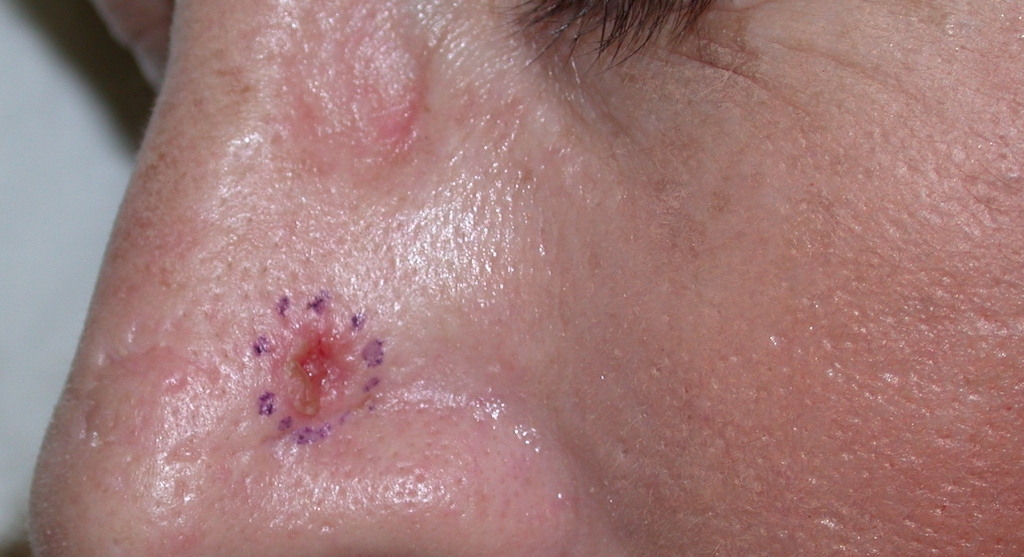
Figure 3. BCC on the nose. Image reproduced with permission of Dr Chris Kearney
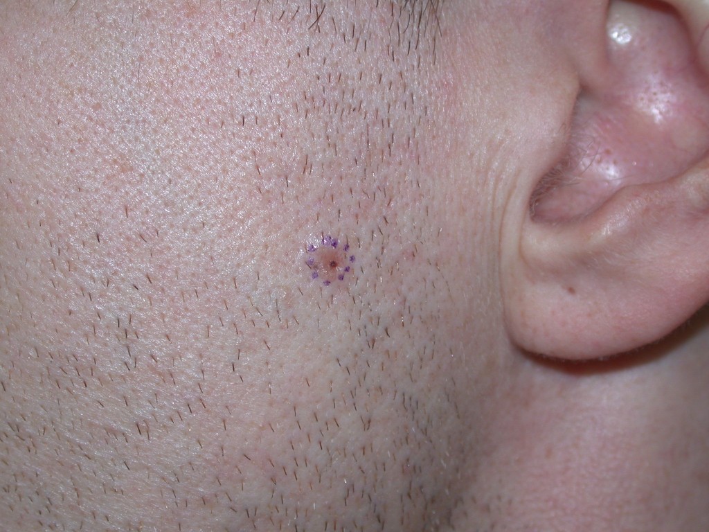
Figure 4. BCC on the cheek. Image reproduced with permission of Dr Chris Kearney
How is basal cell carcinoma diagnosed?
BCC is diagnosed clinically by the presence of a slowly enlarging skin lesion typical appearance. Usually a biopsy (a small sample of the lesion) is taken and sent to the pathology laboratory to be examined under the microscope to confirm the diagnosis.
How is basal cell carcinoma treated?
Treatment options will vary depending on the individual and their needs, and may include:
- Serial cryotherapy
This involves spraying the tumour with liquid nitrogen (-196°c) including a small rim of surrounding skin. The freeze time is prolonged and multiple freeze thaw-cycles may be used, as determined by the treating doctor. This method is mainly used to treat superficial BCCs.
- Standard surgical excision (removal)
This involves cutting out the lesion with a safety margin and suturing (stitching) the skin back together.
- Mohs micrographic surgery
This technique was developed especially for BCCs found on the face where it is important to remove them with a very high cure rate as well as minimise the amount of skin removed in order to achieve the best possible cosmetic outcome. It involves surgical excision (removal) of the lesion with a narrow margin, testing of the pathology while the person waits, ensuring no BCC is left behind, before suturing (stitching) the wound.
- Curettage and cautery
Curettage and cautery involve injection of local anaesthetic, followed by scraping the lesion from the skin surface with a curette and then cauterisation (heat-sealing with a hot wire) of the base. It typically heals with a pale scar a little wider than the lesion that was treated. It is often used for superficial BCCs on the trunk and limbs.
- Photodynamic therapy
This involves application of a sensitising cream followed by exposure to a special red light. It has a lower chance of scar formation than other treatments; however, it is only appropriate for certain superficial and low-grade lesions.
- Topical immunotherapy
Topical immunotherapy involves application of a special cream. The cream stimulates the body’s own immune system to fight and destroy the skin cancer cells. Typically, the area will become moderately inflamed during the period of treatment. It is only appropriate for certain superficial and low-grade lesions.
- Radiotherapy
Radiotherapy is occasionally used if other treatments are not possible. In the case of an advanced BCC, radiotherapy is sometimes used after, and in addition to, surgical excision (removal).
What is the likely outcome of basal cell carcinoma?
Fortunately, BCC is usually the least dangerous and almost always completely curable by treatment. It is extremely rare for a BCC to spread to other parts of the body however they will relentlessly enlarge and destroy nearby tissue if not treated. They have an affinity for nerves, and some can spread along a nearby nerve, making them much more difficult to cure.
After having a single BCC there is, on average, a 50% chance of developing another BCC at some stage of a person’s life. 2
The general recommendation from dermatologists after removing a BCC is to undergo skin-checks every 6 to 12 months. It is important that the skin be self-examined, and any suspicious new lesions reported.
- Bartos V. Development of Multiple-Lesion Basal Cell Carcinoma of the Skin: A Comprehensive Review. Sisli Etfal Hastan Tip Bul. 2019 Nov 19;53(4):323-328. doi: 10.14744/SEMB.2019.08058. PMID: 32377105; PMCID: PMC7192293.
- Karagas MR, Stukel TA, Greenberg ER, Baron JA, Mott LA, Stern RS. Risk of Subsequent Basal Cell Carcinoma and Squamous Cell Carcinoma of the Skin Among Patients With Prior Skin Cancer. JAMA. 1992;267(24):3305–3310. doi:10.1001/jama.1992.03480240067036
| Dr Chris Kearney | December 2023 |
| Dr Chris Kearney | October 2022 |
Disclaimer
2019 © Australasian College of Dermatologists.
You may use for personal use only. Please refer to our disclaimer.